Put your notes in here. Type them, publish them, view them, edit them, save it and print them. Notes are stored locally, if you clear your browser history they will disappear. Notes are a great way to add your own notes, from other sources to this station.
Orthopaedic Skills And Examination
Hip Examination

Introduction
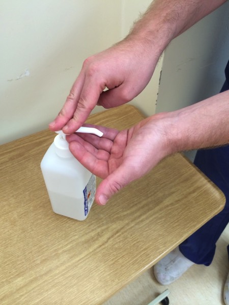
Wash your hands
Introduce yourself to the patient
Explain that you would like to examine their hip joint
Check whether they are in any pain and which the ‘bad’ hip is
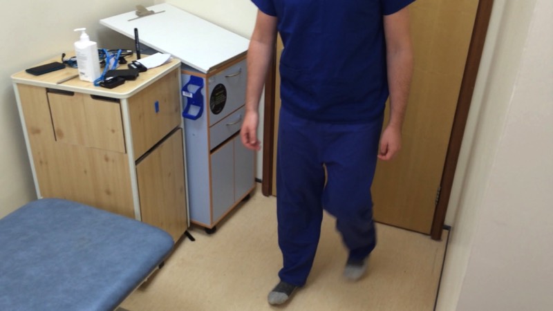
Walk
Asking the patient to walk a short distance is a good screening test and allows you to combine ‘looking’ at the hip and leg, analysing gait and looking for any gross deformity.
Gait
- Antalgic: shorter steps due to pain, the commonest cause is OA.
- Scissor: legs move with a rigid outward swing resembling a pair of scissors . Due to hypertonic legs, associated with UMN lesion particularly cerebral palsy
- Trendelenburg: weakness of abductor muscles. The pelvis sags on the opposite side of the superior gluteal nerve lesion
- Ataxic: unsteady, broad-based gait associated with cerebellar dysfunction.
- High Stepping/Foot drop: due to loss of dorsiflexion secondary to peroneal nerve injury. The patient is unable to dorsiflex and compensates with a high step prior to landing the foot.
Although ‘Special Tests’ are often left till the end of the examination, while you have the patient standing, now is a good time to perform Trendelenberg’s Test to assess abductor function.
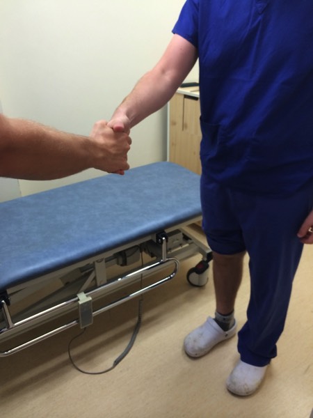
Trendelenburg's Test
- Stand in front of the patient facing them and take their hands in yours. Ask the patient to stand on one leg. Normally the pelvis tilts up on unsupported side (abductors on weight bearing side). A positive Trendelenburg is when the unsupported side droops, indicating pathology on the leg on which they are standing. A mnemonic is: "sound side sags", indicating the normal leg which is in the air sags. Causes: weakness of the abductors, dislocation/fractures, pain, fixed flexion deformity.

Look
Position the patient standing up in their underwear
Check the front and back then position them on the examination couch
- Skin: Scars (check buttocks), swelling, venous/arterial disease.
- Muscle: Wasting of the quads/glutei/hamstrings
- Bone & Joint: Length, deformities (e.g. leg shortened or externally rotated)
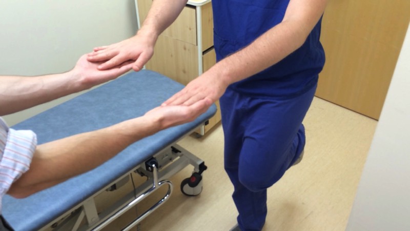
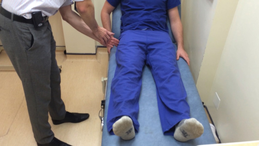
Feel
Measure
Position the pelvis so ASIS at same level
True length: Distance from medial malleolus to ASIS each side
Apparent length: Distance from medial malleolus to bottom of sternum
Feel
Palpate for bony prominences. Greater Trochanter, ASIS, iliac crests, pubic rami. The hip is deep so tenderness is unlikely
Move
Active Movements: The patient moves
Passive Movements: The doctor moves
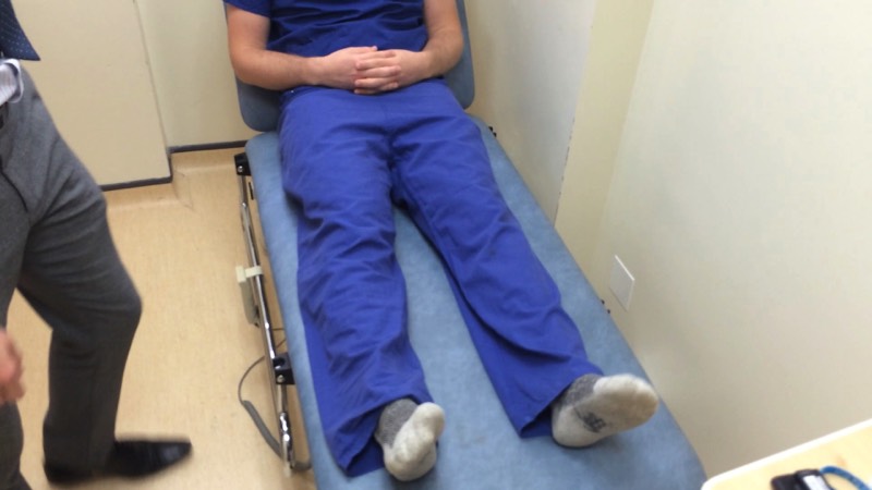
With the patient supine, the hip should be stabilzed by putting your arm across their pelvis.
Compare each side as you go
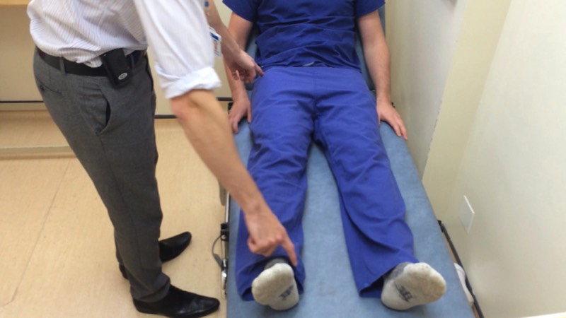
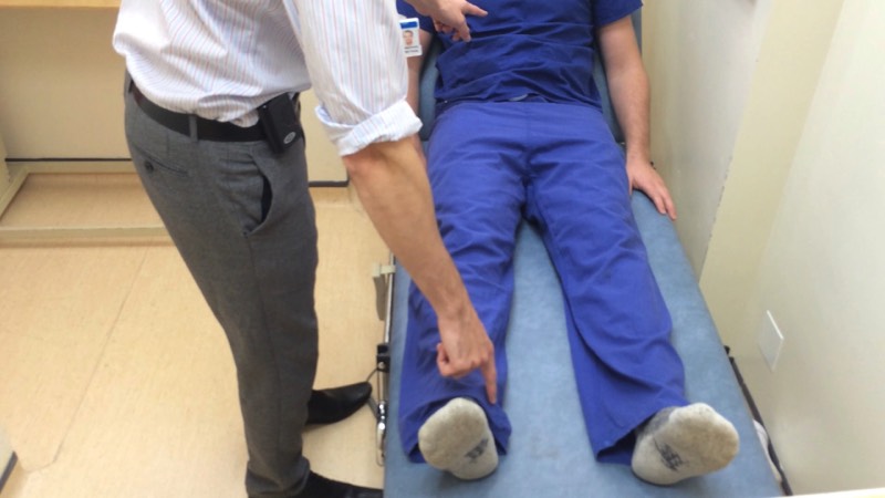
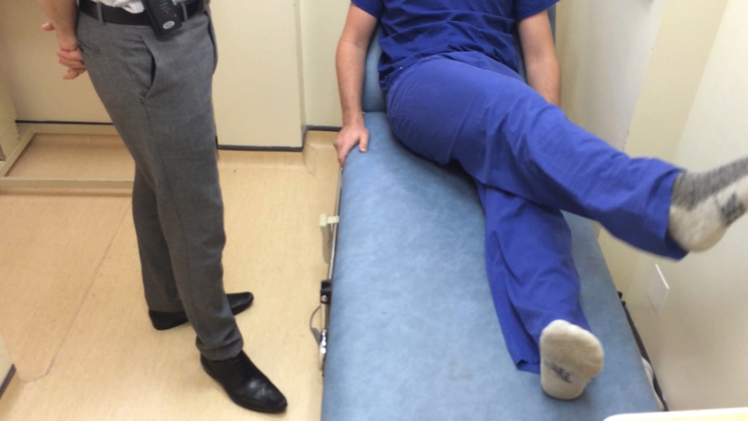
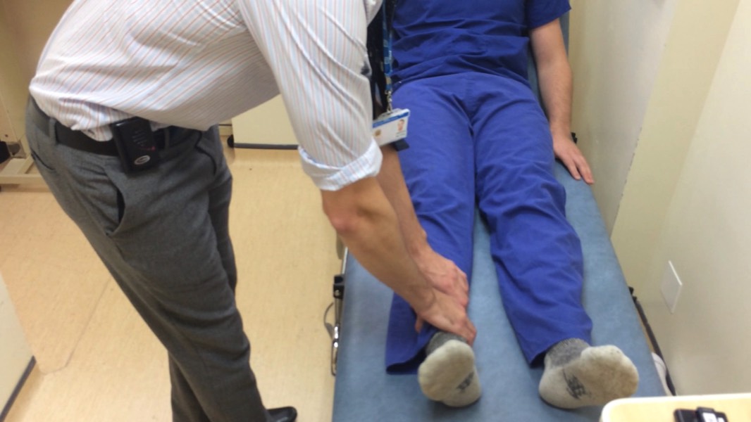
A good screening test for gross hip irritability is internal/external rotation with the leg extended in the supine position
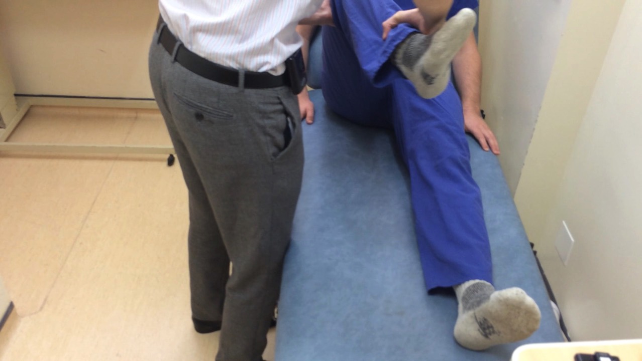
- Flexion (0-120°): bring your knee up to your stomach
- Extension (0-10°): best done with the patient standing or lying prone, move leg backwards.
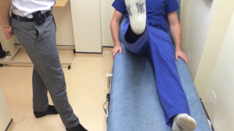
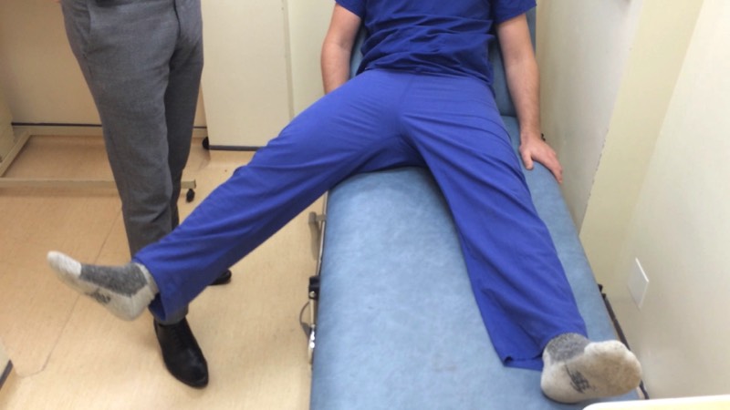
- Abduction (0-45°): with a straight leg move the leg outwards
- Adduction (0-30°): with a straight leg move the leg across to the opposite side
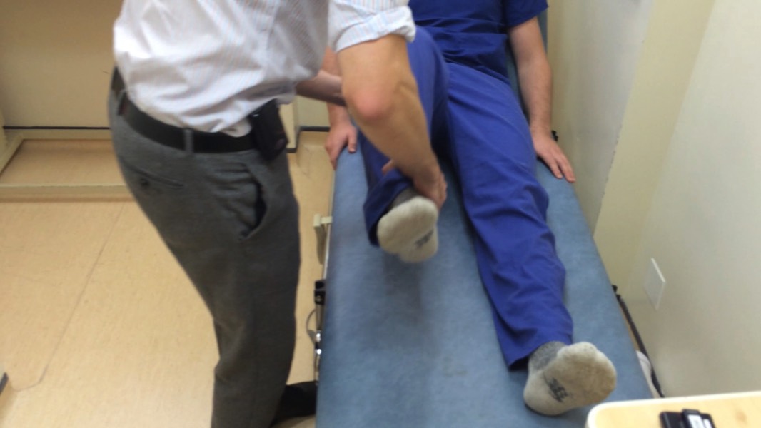
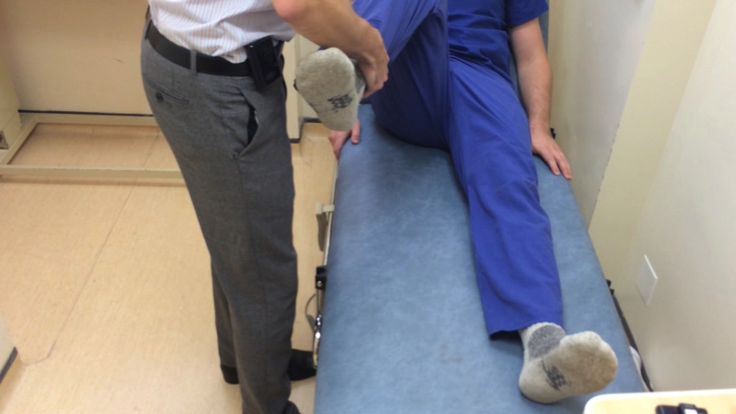
- External Rotation (0-45°): rotate thigh outwards
- Internal Rotation (0-30°). rotate thigh inwards, do this with the knee and hip flexed
Special Tests
Trendelenberg and Gait have been assessed at the beginning of the examination.
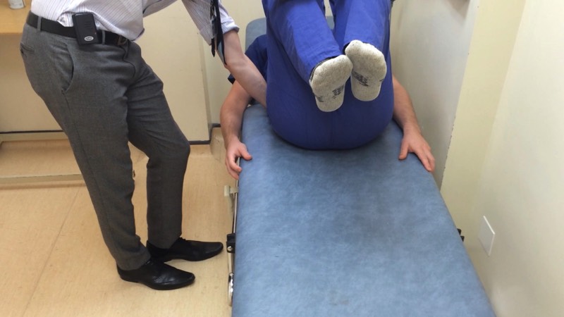
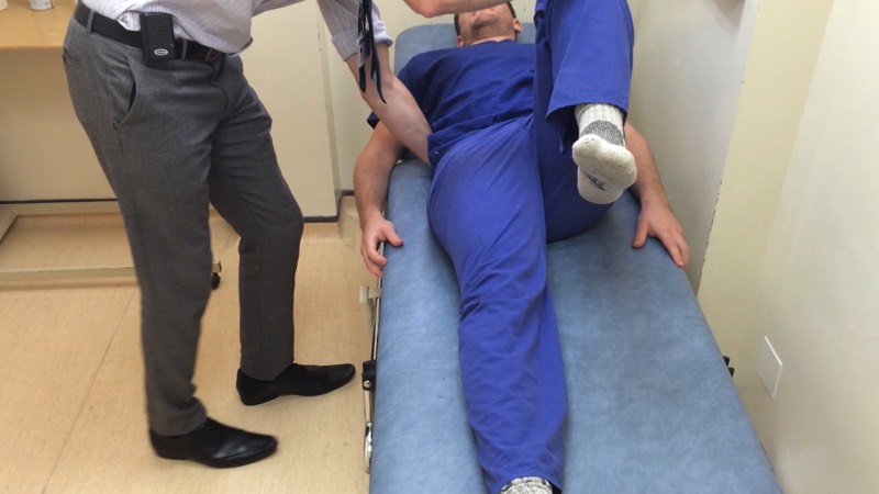
Fixed Flexion (e.g. OA)
- Thomas' test: Fully flex both legs to obliterate lumbar lordosis (can place one hand under back to confirm). Hold one leg fully flexed and straighten the other. The patient should be able to lie the other leg fully on to the bed, any angle left is the degree of fixed flexion. Repeat on other side.
Closing
Thank the patient
Wash your hands
State that you would like to:
Wash your hands
State that you would like to:
- Examine the joint above and below (lumbar spine and knee)
- Get AP and lateral radiographs of the hip
- Assess the patient’s general fitness for surgery
P
Medical imagery licensed under Creative Commons Attribution-Share Alike license; sourced from Wikipedia
All other textual content, imagery, and website design copyright © 2014-22 MRCS Part B Questions all rights reserved.
Contact Us | Privacy Policy | Terms and Conditions